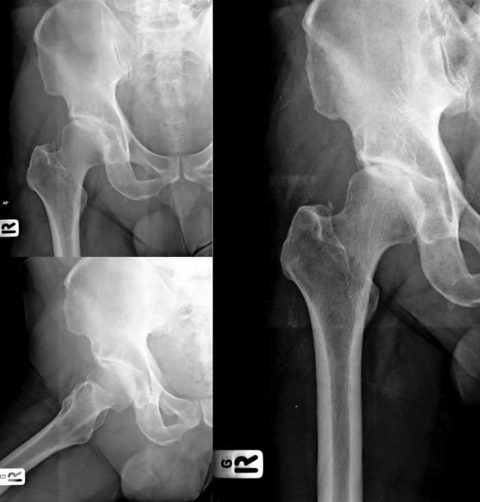
Treatment for hip dysplasia can restore mobility and comfort.
Hip dysplasia is an orthopedic condition in which the patient’s hip socket doesn’t fully cover the ball portion of the femur. This means that the joint is much more likely to dislocate. Additionally, this malformation causes extra stress the joint, which can wear down the cartilage at a faster rate than usual.
What Causes It?
Most patients with hip dysplasia are born with the condition, and it isn’t always fully understood why the patient has this malformation. However, there are some risk factors that increase the likelihood that a baby will have hip dysplasia. For example, hip dysplasia can run in families, although an exact pattern isn’t known. The main risk factors seem to be related to the amount of room the baby has while in the womb.
For this reason, first-born babies and babies that are large seem to be at an increased risk. Babies that are in the breech position just before birth are also more likely to have hip dysplasia because the hip joint isn’t in the proper position to be able to grow. In the womb, the hip joint is actually composed of cartilage, so when there isn’t adequate space for the hip socket to form, it will develop so that it only partially covers the ball. As the person grows, the hip will turn to bone, but the malformation often persists.


What Are The Symptoms?
The symptoms of hip dysplasia aren’t always obvious, particularly because infants can’t communicate them. This is why it is critical for medical staff to evaluate the infant for hip dysplasia both at birth and through development. There are some signs and symptoms that parents can watch out for as their child develops though. Even before the patient is able to communicate, parents may be able to notice if one leg appears longer than the other or if the child favors one leg over the other as they begin to crawl. Typically, the signs will become more obvious as the patient begins to walk. This is when their parents would be able to identify a limp.
If the case is minor, the patient may not notice any signs until they are much older and are participating in activities that require flexibility. The patient will likely report feeling pain either in the groin, side of the hip, or back of the hip. This pain tends to gradually become more noticeable as the cartilage wears down. Patients may also experience frequenting popping in the joint.
How Is It Diagnosed?
Screening for hip dysplasia is routine in infants, so the infant’s physician should test for it by checking the range of motion of the hips. If the physician has reason to believe that the infant may have it, such as the baby being in breech position, they will likely order an ultrasound to confirm the diagnosis. If the symptoms don’t appear until late in life, the physician will order MRIs to test for cartilage wear, and an X-ray to confirm suspected hip dysplasia.
What Are The Treatment Options?
Catching hip dysplasia in the infant stage can help prevent damage over the years and make the treatment process less complicated. In many infant cases, the joint will be placed in a cast to allow the hip to grow properly around the ball joint. However, some infant cases will require a full-body cast. If the malformation isn’t caught until later in life, the patient will likely have to undergo surgery to make the hip fit around the ball joint properly.
